Upper Back Anatomy / Upper Back Pain Local Physio : Both the deltoid and the trapezius are firmly attached to the spine of the scapula.. The back functions are many, such as to house and protect the spinal cord, hold the body and head upright, and adjust the movements of the upper and lower limbs. The human spine is composed of 4 sections of vertebrae. Human muscles · may 25, 2020. The intricate anatomy of the back provides support for the head and trunk of the body, strength in the trunk of the body, as well as a great deal of flexibility and movement. The cervical spine protects the nerves connecting to.
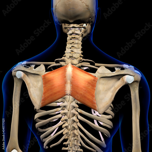
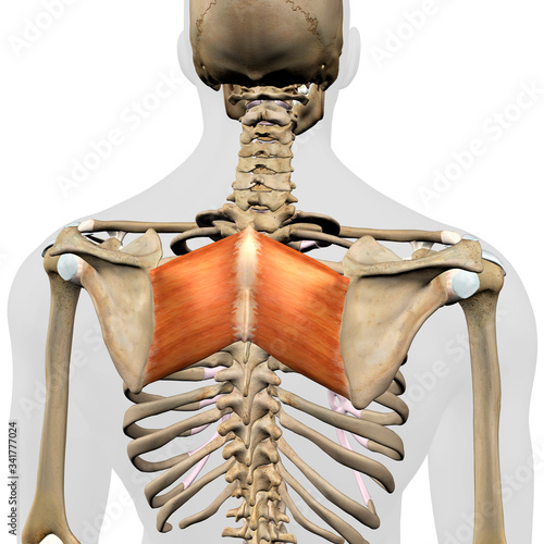
The rib cage also anchors the bones of the head, neck, shoulders, and arms to the trunk of the body. The upper back muscles of the rhomboids and the trapezius are responsible for many of the movements of the scapula which in turn plays a huge role in the stability and mobility of the shoulder. Human muscles · may 25, 2020. Anatomy of the upper back muscles. The trapezius and latissimus dorsi muscles connect the upper limb to the vertebral column.
Rhomboid Major Muscles In Isolation Rear View Of Upper Back Human Anatomy Wall Mural Hank Grebe from t3.ftcdn.net Both the deltoid and the trapezius are firmly attached to the spine of the scapula. The iliocostalis muscles are furthest from the spine. Upper back pain is most commonly caused by muscle irritation or tension, also called myofascial pain. Back anatomy the back is the body region between the neck and the gluteal regions. The rib cage also anchors the bones of the head, neck, shoulders, and arms to the trunk of the body. It consists of seven vertebrae. It runs from the neck to the upper back. The neck consists of seven cervical vertebrae, the building blocks of the spine.
The iliocostalis muscles are furthest from the spine.
The bones of the chest and upper back combine to form the strong, protective rib cage around the vital thoracic organs such as the heart and lungs. Human muscles · may 25, 2020. The thoracic spine —also referred to as the upper back or middle back—is designed for stability to anchor the rib cage and protect vital internal organs within the chest. This muscle is located on the upper portion of the back anatomy, underneath the trapezius. It comprises the vertebral column (spine) and two compartments of back muscles; The nervous system of the thorax is a vital part of the nervous system as a whole, as it includes the spinal cord, peripheral nerves, and autonomic ganglia that communicate with and control many vital organs. The thigh bears much of the load of the body's weight when a person is upright. It is like that for several reasons, all of which you can understand by looking at the anatomy of the thoracic spine. The extrinsic (superficial) back muscles, which lie most superficially on the back. The upper back has the most structural support, with the ribs attached firmly to each level of the thoracic spine and very limited movement. They originate from the vertebrae and insert into the scapulae. The basic anatomy of your upper back by lindsey mcfadden as you're doing your regular upper back stretching exercises , you're probably wondering about the components of your upper back and why it happens to be the most stable part of your spine. The cervical spine supports the weight and movement of your head and protects the nerves exiting your brain.
The nervous system of the thorax is a vital part of the nervous system as a whole, as it includes the spinal cord, peripheral nerves, and autonomic ganglia that communicate with and control many vital organs. Human body anatomy female female anatomy muscle shoulder blade pain anatomy back muscles bones man female anatomy body muscles in a body female anatomy muscole shoulder concept muscular sysyem. The muscles of the back are a group of strong, paired muscles that lie on the posterior aspect of the trunk they provide movements of the spine, stability to the trunk, as well as the coordination between the movements of the limbs and the back muscles are divided into two large groups: The human spine is composed of 4 sections of vertebrae. Back muscles anatomy here include the trapezius, latissimus dorsi, rhomboid and levator scapulae.
Rhomboid Major Muscles In Isolation Rear View Of Upper Back Human Anatomy Wall Mural Hank Grebe from t3.ftcdn.net The spine is made up of 33 individual bones called ve. The cervical spine is the top part of the spine. The muscles of the back are a group of strong, paired muscles that lie on the posterior aspect of the trunk they provide movements of the spine, stability to the trunk, as well as the coordination between the movements of the limbs and the back muscles are divided into two large groups: It comprises the vertebral column (spine) and two compartments of back muscles; Vertebrae there are 12 vertebrae in the thoracic spine. The trapezius and latissimus dorsi muscles connect the upper limb to the vertebral column. Powerful muscles that move the head and arms attach to these bones as well. It is very stiff, and the thoracic spine has a limited range of motion.
The rib cage also anchors the bones of the head, neck, shoulders, and arms to the trunk of the body.
It comprises the vertebral column (spine) and two compartments of back muscles; The thigh bears much of the load of the body's weight when a person is upright. It is very stiff, and the thoracic spine has a limited range of motion. Upper back pain rear view of spine back pain spine sports spine spine surgery spine white background back ache x human anatomy illustration human anatomy on white background upper body stretch. Both the deltoid and the trapezius are firmly attached to the spine of the scapula. See human back anatomy stock video clips. See upper back stock video clips. The back functions are many, such as to house and protect the spinal cord, hold the body and head upright, and adjust the movements of the upper and lower limbs. The cervical spine protects the nerves connecting to. The rhomboid muscle is activated as you bring and squeeze your scapula or shoulder blades back and together. The seventh cervical vertebra, referred to as c7, meets the first of 12 thoracic vertebrae t1 at the base of the neck, a. The bones of the chest and upper back combine to form the strong, protective rib cage around the vital thoracic organs such as the heart and lungs. It contains many muscles and nerves but only has one bone, the femur, which is the longest and strongest bone in.
Upper back pain rear view of spine back pain spine sports spine spine surgery spine white background back ache x human anatomy illustration human anatomy on white background upper body stretch. It is like that for several reasons, all of which you can understand by looking at the anatomy of the thoracic spine. The trapezius and latissimus dorsi muscles connect the upper limb to the vertebral column. Before giving our recommendations for upper back exercises, it's important to first go over the anatomy of the back musculature. The main superficial muscles of the back are the following:
1 from Upper back pain is most commonly caused by muscle irritation or tension, also called myofascial pain. The cervical spine protects the nerves connecting to. It is like that for several reasons, all of which you can understand by looking at the anatomy of the thoracic spine. This is my video about the muscles of the back. See human back anatomy stock video clips. Anatomy of the upper back muscles. The rib cage also anchors the bones of the head, neck, shoulders, and arms to the trunk of the body. The upper back has the most structural support, with the ribs attached firmly to each level of the thoracic spine and very limited movement.
Each block is separated by a disc that sits in between and each vertebra has a facet joint on either side.
It is like that for several reasons, all of which you can understand by looking at the anatomy of the thoracic spine. The rhomboid muscle is activated as you bring and squeeze your scapula or shoulder blades back and together. Anatomy of the upper back muscles. The seventh cervical vertebra, referred to as c7, meets the first of 12 thoracic vertebrae t1 at the base of the neck, a. The deltoid, teres major, teres minor, infraspinatus, supraspinatus (not shown) and subscapularis muscles (not shown) all extend from the scapula to the humerus and act on the shoulder joint. The upper back muscles of the rhomboids and the trapezius are responsible for many of the movements of the scapula which in turn plays a huge role in the stability and mobility of the shoulder. It comprises the vertebral column (spine) and two compartments of back muscles; The trapezius and latissimus dorsi muscles connect the upper limb to the vertebral column. Anatomy of the back organs. Human body anatomy female female anatomy muscle shoulder blade pain anatomy back muscles bones man female anatomy body muscles in a body female anatomy muscole shoulder concept muscular sysyem. The human spine is composed of 4 sections of vertebrae. These sections are cervical (neck), thoracic (upper and middle back), lumbar (lower back), and sacrum (tailbone). This muscle is located on the upper portion of the back anatomy, underneath the trapezius.


0 Komentar